Review the six linked lung ultrasound images labelled A to F.
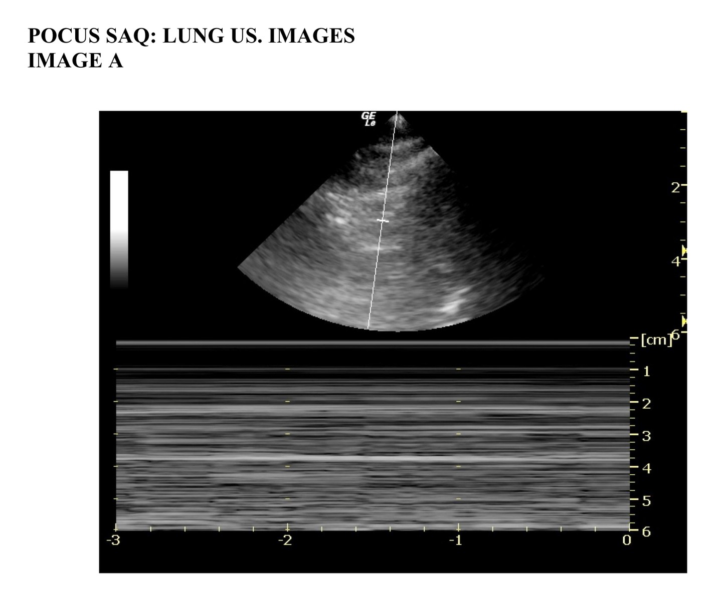 Image A
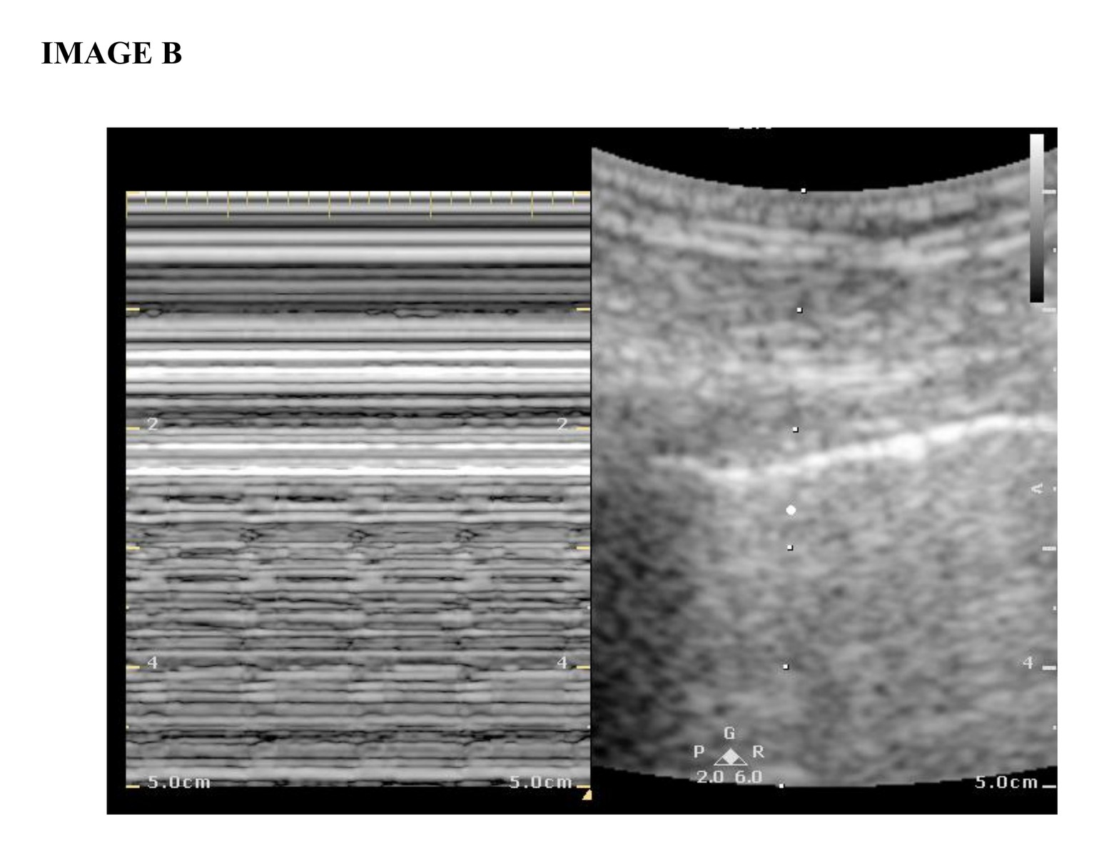 Image B
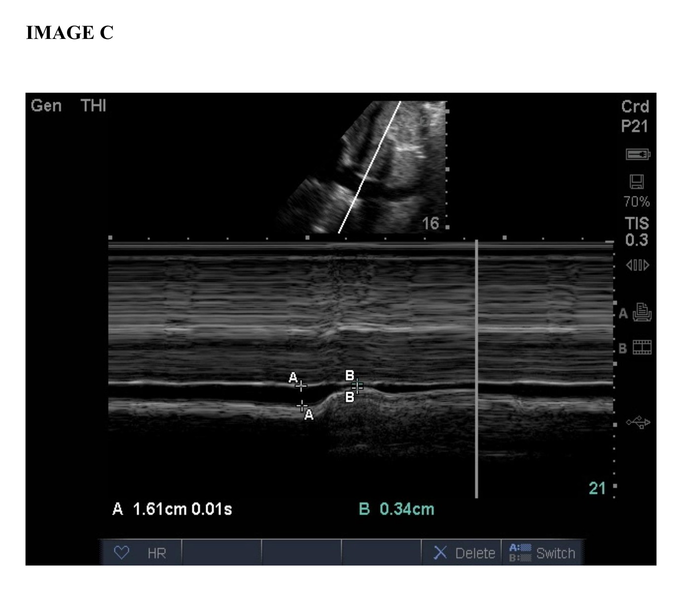 Image C
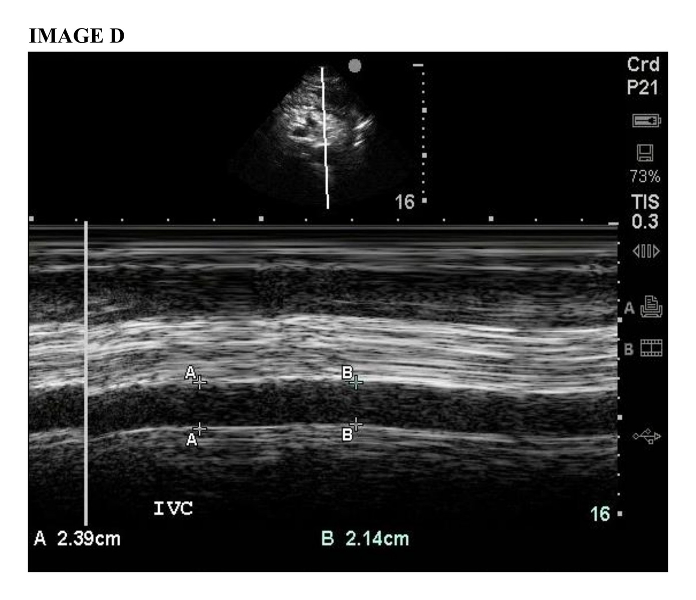 Image D
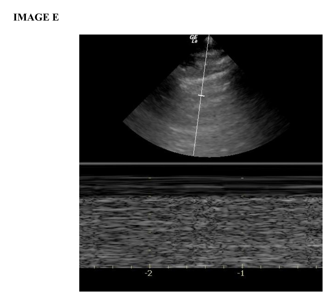 Image E
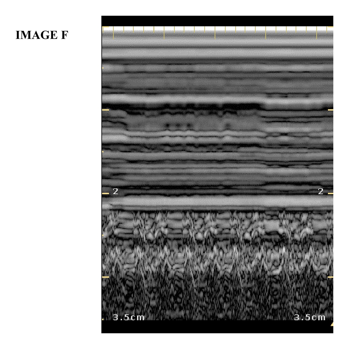 Image F
Which M-mode image, A or B, demonstrates pneumothorax?
(Marked out of 1.0)
/ 1
Explain why the other image is not consistent with pneumothorax.
(Marked out of 4.0)
/ 4
Which M-mode IVC image, C or D, is consistent with tension pneumothorax physiology?
(Marked out of 1.0)
/ 1
Explain why the other IVC image is not consistent with tension pneumothorax.
(Marked out of 4.0)
/ 4
Which image, E or F, is consistent with pleural sliding?
(Marked out of 1.0)
/ 1
Explain why the other image is not consistent with pleural sliding.
(Marked out of 4.0)
/ 4
Total Score: 0 / 15
Percentage: 0%
Comments are closed for this SAQ.