A 25-year-old woman is brought to the ED after a high-speed head-on motor vehicle crash. Her initial chest X-ray and a later CT head slice are shown.
After the chest injury has been treated, she is intubated for assessment and management. The ventilator alarms for high pressures. CT has shown only the chest and head injuries in the images. She has a dilated left pupil. Her vital signs are GCS 3 sedated and paralysed, pulse 100/min, BP 90/60 mmHg, and oxygen saturation 90% on FiO2 0.5.
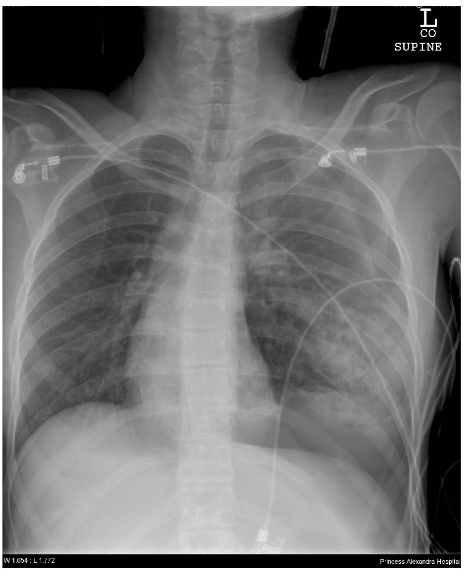 Chest X-ray.
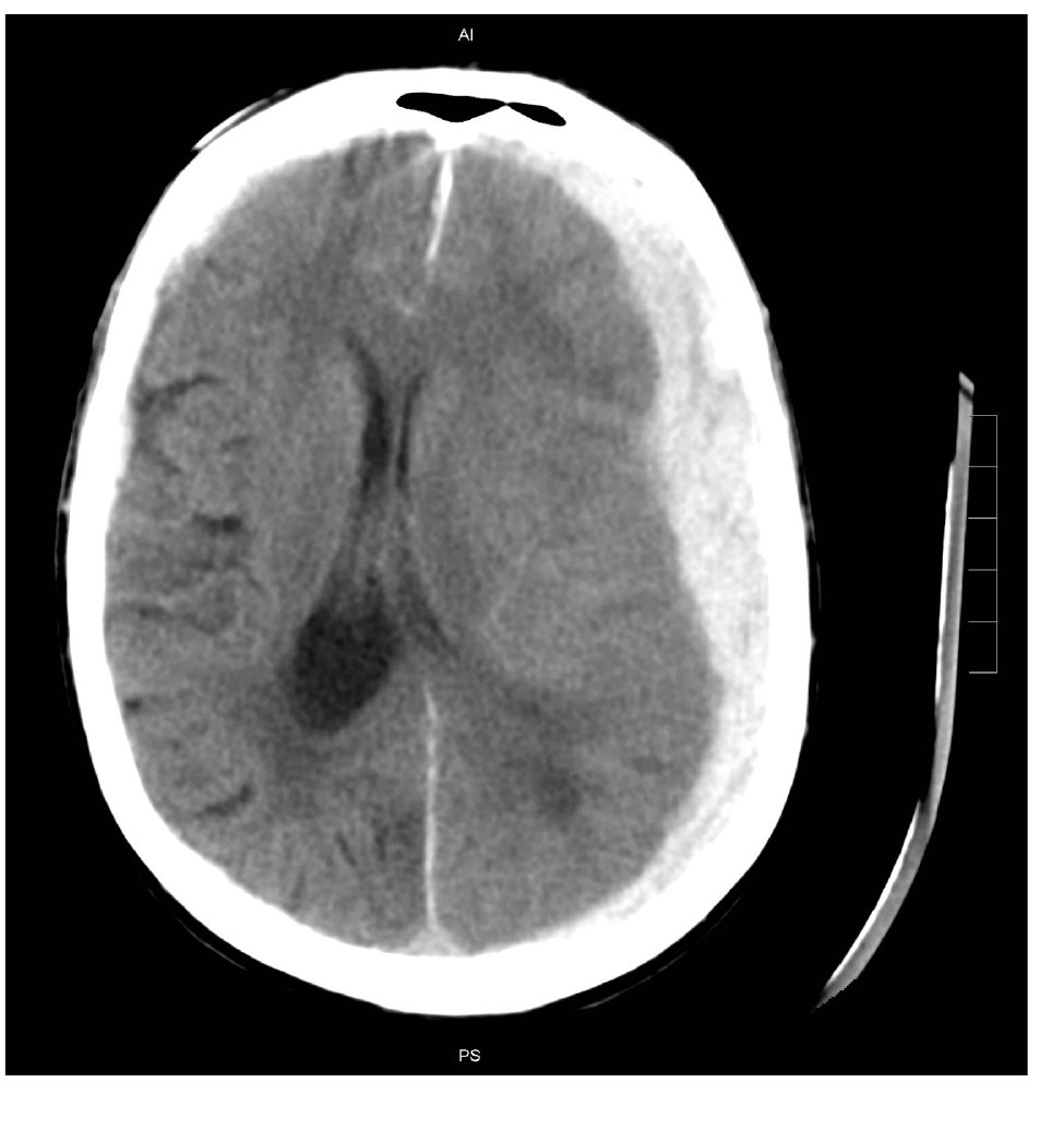 CT head.
Give the most important pathology on the chest X-ray, with three radiographic supporting features.
(Marked out of 4.0)
/ 4
State the definitive treatment for the diagnosis in part A.
(Marked out of 1.0)
/ 1
State the other clinically significant chest X-ray pathology and its radiographic support.
(Marked out of 2.0)
/ 2
List six possible causes of high ventilator pressures.
(Marked out of 6.0)
/ 6
List the abnormalities on the CT head image.
(Marked out of 6.0)
/ 6
State your management steps, including drugs, doses, and treatment endpoints where appropriate.
(Marked out of 8.0)
/ 8
Total Score: 0 / 27
Percentage: 0%
Comments are closed for this SAQ.